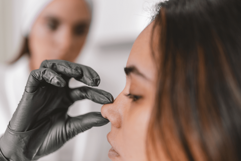
How is Rhinoplasty (Nose Job) Surgery Performed?
Rhinoplasty is a highly personalized surgical procedure that aims not only to change the external appearance of the nose but also to improve respiratory functions. The surgery is typically performed under general anesthesia in a sterile operating room environment of a fully equipped hospital and lasts an average of 2 to 4 hours.
1. Preparation and Anesthesia
Before the surgery, the patient’s general health status is evaluated by an anesthesiologist. Rhinoplasty is almost always performed under general anesthesia to ensure the patient’s comfort throughout the surgical process. After the patient is asleep, the surgical team completes the sterilization procedures and begins the operation.
2. Selection of Incision Technique
Rhinoplasty is fundamentally performed using two main techniques:
- Open Rhinoplasty: In this technique, a small, inverted “V” incision is made on the columella, the external part separating the nostrils. This incision allows the nasal skin to be lifted and the surgeon to directly visualize and work on the nasal skeletal structure (bone and cartilage). It is preferred for correcting complex deformities, tip shaping, and severe asymmetries.
- Closed Rhinoplasty: All incisions are made inside the nostrils. The surgical field of view is more limited, but there is no visible external scar. It may be suitable for smaller corrections and simpler cases.
Today, open rhinoplasty technique is more commonly used because it provides the surgeon with greater control.
3. Shaping the Cartilage and Bone Structure
The most critical stage of the surgery is the reshaping of the nasal skeleton:
- Dorsal Reduction (Hump Reduction): If desired, the dorsal hump on the bridge of the nose is carefully removed or filed down. This procedure is often performed with modern, controlled instruments like piezo surgery (ultrasonic bone cutting) to minimize trauma and bruising.
- Nasal Tip Shaping (Tip Plasty): The cartilages that form the nasal tip (alar cartilages) are reshaped. The surgeon uses sutures and small pieces of cartilage taken from the nasal septum or ear (grafts) to give the nasal tip projection, definition, and symmetry.
- Narrowing the Wide Nasal Bridge (Osteotomy): If the nasal bones remain excessively wide after the dorsal hump is removed, controlled cuts (osteotomy) are made in the nasal bones, and the bones are moved inward to slim the nasal bridge and create an aesthetic transition.
- Septum Correction (Septoplasty): Deviations in the nasal septum, which cause breathing problems, are corrected simultaneously.
4. Use of Cartilage Grafts
Cartilage grafts are vital to support the nose’s new structure and ensure long-term stability. These grafts are most often taken from the patient’s own nasal septum, and sometimes from the ear or rib cartilage, and used to strengthen the bridge (strut grafts) or provide support to the tip.
5. Closure and End of Surgery
Once the nose has achieved the desired shape, all incisions (the columella incision in open rhinoplasty) are closed with very fine sutures. Silicone splints or thin splints may be placed inside the nose to support the internal structures and prevent adhesions. Finally, a thermoplastic splint (cast) or special tapes are applied to the nasal bridge to protect the external structure and reduce swelling. The patient is taken to the recovery room to wake up and is typically discharged after being monitored in the hospital overnight.
Day-by-Day Breakdown of the Process
Day 1: Arrival in Turkey – VIP transfer to hotel, rest and relaxation.
Day 2: Consultation with the surgeon and all necessary tests.
Day 3: Surgery Day
Day 3: Post-operative examinations and check-ups continue.
Day 4: Enjoy free time to explore Istanbul before your transfer to the airport (optional).
How to Book Your Hair Transplant with Cure Holiday?
- Step 1: Contact us for your free consultation and receive a personalized treatment plan.
- Step 2: Choose your preferred dates and confirm your booking.
- Step 3: Arrive in Turkey, and let us handle the rest!
All-Inclusive Rhinoplasty Surgery
Explanation of Rhinoplasty Techniques
The name of the technique applied in rhinoplasty operations is determined by how the surgeon accesses the internal structure of the nose. There are two main surgical approaches: Open Rhinoplasty and Closed Rhinoplasty.
1. Open Rhinoplasty
In the Open Rhinoplasty technique, a small, horizontal incision is made across the strip of tissue separating the nostrils (the part called the columella). This incision allows the nasal skin to be lifted upwards, providing a wide and direct view of the underlying cartilage and bone structures.
- Advantages: It enables the surgeon to see all the structures clearly, allowing for more complex corrections (advanced deformities, shaping with cartilage grafts, or revision surgeries) to be performed with greater precision.
- Disadvantages: A small, externally visible scar remains on the columella area, which usually becomes inconspicuous after healing.
2. Closed Rhinoplasty
In the Closed Rhinoplasty technique, all surgical incisions are made inside the nostrils. There are no external visible scars. The surgeon works through a narrow field, shaping the bone and cartilage structures from within, without lifting the nasal tip.
- Advantages: The biggest advantage is the absence of external visible scars. According to some surgeons, swelling and recovery time may potentially be faster.
- Disadvantages: The surgeon’s working area and field of view are more limited compared to Open Rhinoplasty. Therefore, it is generally preferred for simpler cases or those requiring limited correction.
Additional Technique: Piezo (Ultrasonic) Rhinoplasty
Piezo Rhinoplasty (or Ultrasonic Rhinoplasty), which has gained popularity in recent years, refers to the method of shaping the bones, independent of the access techniques mentioned above (Open or Closed). In this technique, bone cutting and shaving are performed using special devices (piezoelectric surgical instruments) that generate ultrasonic vibrations, instead of traditional tools.
- Purpose: Since ultrasonic waves primarily affect the bone, the risk of damage to the surrounding soft tissues, cartilage, vessels, and nerves is lower compared to traditional methods. This can help reduce bruising and swelling after the surgery.

