
Part of: Eye treatment in Turkey — prices, what is included and country comparison.
Cataracts are the most common cause of vision loss and blindness worldwide. This condition, which naturally occurs as a result of aging, is characterized by the eye’s natural lens gradually losing its transparency and becoming increasingly cloudy. Cataract treatment, once considered complex and risky, has become an extremely safe, effective, and routine procedure with advances in modern medicine.
Cataract surgery now not only corrects blurred vision but also has the potential to fundamentally improve patients’ quality of life by simultaneously addressing astigmatism, near, and distance vision problems. This comprehensive guide deeply explores all the details of the treatment journey, from what a cataract is to the most advanced surgical methods, intraocular lens options, and the recovery process.
What is a Cataract and Why is Treatment Necessary?
The natural lens of the eye, located just behind the iris and pupil, is a clear structure responsible for focusing light onto the retina. The formation of a cataract means that the proteins making up this lens begin to deteriorate, clump together, and the lens loses its clarity. This causes light entering the field of vision to scatter, leading to blurriness and fading colors.
Most cataract cases are age-related (senile cataracts), but they can also occur due to secondary causes such as diabetes, long-term steroid use, eye injuries, or genetic factors. Although the progression of a cataract varies depending on its type and the patient’s general health, it is a progressive process and does not regress on its own.
Intervention is necessary when the cloudy lens starts to severely affect daily life, similar to looking through a dirty window. If left untreated, cataracts can progress to severe vision loss that prevents a person from performing daily activities (driving, reading, recognizing faces). Furthermore, very hardened cataracts (mature or hypermature cataracts) can make surgery technically more difficult and lead to secondary problems like glaucoma.
The primary goal of treatment is to restore clear vision and thus the independent quality of life by removing the opaque natural lens and replacing it with a clear, artificial intraocular lens (IOL). Current data shows that cataract surgery is one of the surgical procedures with the highest success and safety rates.
What are the Symptoms of a Cataract and When Should Surgery Be Considered?
A cataract is a slow-progressing disease, and symptoms often start subtly. Patients typically notice a progressive and insidious clouding that begins in one eye. The most common symptom is blurry and hazy vision, which feels like looking through a foggy or frosted pane of glass. This blurriness does not clear up by cleaning glasses or trying a new prescription. Other common symptoms include:
- Difficulty with Night Vision: Seeing halos or glare around oncoming headlights or streetlights, especially when driving at night, which makes nighttime driving hazardous. As light scatter increases, the ability to see in low-light conditions significantly diminishes.
- Fading of Colors: Colors appear more yellowish, faded, or brownish than before. Patients often realize how vibrant the true colors of the world are only after the surgery.
- Double Vision (Diplopia): Rarely, monocular diplopia (seeing two images with one eye) can be an early symptom of a cataract.
- Frequent Changes in Eyeglass Prescription: Nuclear cataracts, in particular, can cause a temporary increase in myopia due to hardening of the lens center, which may lead to a temporary improvement in near vision (“second sight”), but this rapidly fades and leads to permanent visual impairment.
- Light Sensitivity (Photophobia): Increased sensitivity to bright light.
The right time for surgery depends on how much the cataract affects visual function. Ophthalmologists use visual acuity tests and a comprehensive eye exam to determine the level of the cataract. The only critical criterion is not just that visual acuity drops below a certain level.
The decision for surgery is based on how severely the cataract impacts the patient’s quality of life (driving, reading, working, hobbies). If the cataract seriously impedes these essential activities, the time for surgery has come. Performing surgery earlier prevents the lens from becoming excessively hard, reducing surgical risks, and providing the patient with clear vision comfort sooner.
How is a Cataract Diagnosed?
A cataract is diagnosed with a detailed and comprehensive eye examination by an ophthalmologist. This examination aims to assess not only the presence of the cataract but also its grade, type, and the overall health of the eye.
- Visual Acuity Test: The patient’s distance and near vision capabilities are objectively measured using the standard Snellen chart.
- Pupil Dilation (Dilatation): Dilation of the pupils using special drops is essential for diagnosis. This allows the doctor to see the retina, macula, and optic nerve at the back of the eye more clearly, helping to rule out causes of vision loss other than the cataract (macular degeneration, glaucoma damage).
- Biomicroscopy (Slit Lamp Examination): The doctor uses a special microscope called a slit lamp to examine the anterior segment of the eye and, specifically, the lens at high magnification. This examination helps determine the cataract’s type (nuclear, cortical, posterior subcapsular) and density based on the location of the opacity. These details are important for planning the surgical technique.
- Tonometry (Intraocular Pressure Measurement): Intraocular pressure is measured to detect co-existing conditions such as glaucoma (eye pressure).
- Biometry and IOL Calculations (Surgical Planning): After diagnosis and during the surgical planning phase, a highly sensitive test called biometry is performed. This test uses non-contact optical devices (e.g., IOLMaster, Lenstar) to measure the axial length of the eye (front-to-back length) and the curvatures of the cornea (K-values). This data is vital for calculating the power of the artificial Intraocular Lens (IOL) to be implanted, with millimeter-level precision. The accuracy of the lens calculations is crucial for achieving the goal of spectacle independence after surgery.
- Retina Evaluation (OCT and Ultrasound): Additional tests, such as Optical Coherence Tomography (OCT) or intraocular ultrasound for very advanced cataracts, may be requested to obtain further information about retinal health.
How Does the Pre-Surgery Preparation Process Work?
Although cataract surgery is usually a 15–30 minute, outpatient procedure, the pre-surgical preparation process must be meticulously managed for successful outcomes.
- Systemic Health Check: The patient’s general health status (especially diabetes, hypertension, heart disease) is evaluated in detail. The surgeon must be aware of all medications and supplements the patient is taking. Blood-thinning medications (aspirin, coumadin, etc.) may need to be temporarily discontinued before surgery, with the decision made in coordination with the patient’s cardiologist.
- Final Intraocular Lens Decision: The type of IOL (Monofocal, Toric, Trifocal/EDOF) is finalized based on biometry measurements and the patient’s lifestyle expectations (desire for glasses-free reading, need for astigmatism correction, frequency of night driving). This is the most critical step determining patient satisfaction after surgery.
- Ocular Surface Optimization: Conditions affecting the eye surface, such as dry eye syndrome, eyelid inflammation (blepharitis), or allergies, can affect IOL calculations and increase the risk of infection. These conditions are treated with drops or special hygiene regimens before surgery to optimize the eye surface.
- Pre-Operative Drop Treatment: Some surgeons may recommend starting antibiotic or anti-inflammatory drop treatment one or two days before surgery to minimize the risk of infection and inflammation.
- Day-of-Surgery Instructions: On the morning of the surgery, no makeup or perfume should be worn, and comfortable, easily removable clothing should be chosen. Attention should be paid to having a very light breakfast or fasting, according to the surgeon’s instructions.
Are There Non-Surgical Options for Cataract Treatment?
Unfortunately, there is no scientifically proven and approved permanent non-surgical treatment for cataracts. Since a cataract is a change in the protein structure of the lens that is irreversible and causes opacity, it is impossible to restore vision permanently without physically removing this opaque mass from the eye.
- Current Research: Research is ongoing worldwide into drops aimed at slowing or reversing cataract formation, but none of these drops have yet achieved the efficacy and safety evidence required for routine clinical use.
- Glasses and Lighting: When a cataract is in a very early stage and vision loss is minimal, the following methods can be used to delay surgery:
- A stronger eyeglass prescription or bifocal/multifocal lenses can temporarily increase visual acuity.
- Brighter lighting and magnifiers can offer temporary solutions for reading and near tasks.
However, these methods only alleviate symptoms and do not stop the progression of the cataract. When the cataract progresses, and corrected visual acuity drops below legal driving limits or severely restricts the patient’s daily independence, the only permanent and definitive solution is surgery. Unnecessarily delaying surgery only increases the duration and risk of the surgical procedure due to lens hardening.
Phacoemulsification (Phaco) Method: The Gold Standard Surgery
Today, the Phacoemulsification or simply Phaco method is accepted as the “gold standard” and the most commonly performed technique for cataract surgery. This microsurgical method offers rapid recovery and high precision for patients.
Detailed Steps of the Procedure:
- Micro-Incision: A small tunnel incision, usually less than 2-3 mm, that does not require stitches (self-sealing) is created at the edge of the cornea. Additional micro-incisions (LRI) may be made to correct corneal astigmatism if needed.
- Capsulorhexis: A perfectly circular, smooth, and centered opening (capsulorhexis) is created on the anterior surface of the capsule membrane surrounding the cataractous lens, with the help of special forceps or a laser (if FLACS is used). The quality of this opening is vital for the stabilization and optical performance of the IOL.
- Phacoemulsification: A very fine probe (tip) from the Phaco device is inserted into the eye through the incision. This probe emits high-frequency ultrasonic vibrations to gently break up (emulsify) the hardened cataractous lens into small pieces, and simultaneously vacuums (aspirates) these fragments out of the eye. Modern Phaco devices can automatically adjust the ultrasonic energy according to the hardness of the lens.
- Cortical Cleanup: After the lens nucleus is removed, soft lens remnants (cortex) adhering to the walls of the capsular bag are completely cleaned using a special irrigation and aspiration device (I/A probe). A smooth inner surface of the bag helps reduce the risk of “secondary cataract” (PCO).
- Intraocular Lens (IOL) Implantation: Once the capsular bag is completely clean, a foldable artificial IOL is injected into the bag using a special injector system. The lens unfolds inside the eye and is placed into its permanent position within the posterior, intact capsular membrane.
The minimally invasive nature of Phaco surgery accelerates post-operative recovery and allows the patient to quickly return to their normal life.
What is Femtosecond Laser-Assisted Cataract Surgery (FLACS)?
Femtosecond Laser-Assisted Cataract Surgery (FLACS) is an advanced technology that enhances the precision and reproducibility of cataract surgery. Some critical steps of traditional Phaco surgery are performed not by the surgeon’s manual skill but by a computer-controlled laser firing pulses at one quadrillionth of a second.
Key Steps Performed by FLACS:
- Laser Incisions: Corneal incisions are automatically created by the laser at a pre-planned depth, width, and angle. This ensures more precise placement than manual incisions and can minimize the induction of surgical astigmatism.
- Perfect Capsulorhexis: The laser creates a perfectly circular, well-centered, and precisely sized opening (capsulorhexis) in the capsule membrane surrounding the cataractous lens, far superior to the manual tearing (capsulorhexis) performed by hand. This precision is vital for premium (multifocal) lenses to be perfectly centered on the optical axis of the eye and perform optimally.
- Lens Softening (Nuclear Fragmentation): The laser is used to pre-fragment and soften the hard cataractous lens by creating a grid, star, or cube pattern inside the lens before the use of ultrasound. This pre-fragmentation significantly reduces the amount of ultrasonic energy and time required during the Phaco stage to emulsify the cataract. The reduced use of ultrasonic energy lowers thermal and mechanical stress on intraocular tissues (especially the corneal inner layer), which is important for patients with sensitive corneas or low endothelial cell counts.
FLACS also offers extra precision in determining the axis for Toric lenses. The laser can mark the correct axis where the Toric lens should be placed during surgery. While FLACS is an option, standard Phaco surgery performed by an experienced surgeon is also extremely safe and successful; FLACS is usually preferred in cases where premium lenses are implanted or when extra precision is required for special circumstances.
What Happens During Surgery?
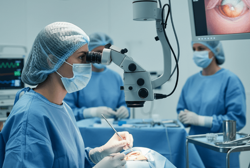
Cataract surgery is a much more comfortable and quicker process than most patients imagine. The procedure is performed in a sterile operating room under a surgical microscope.
- Positioning and Preparation: The patient is taken to the operating table and lies down in a comfortable, supine position. The eye area to be operated on is cleaned with an antiseptic solution and covered with a sterile drape, leaving only the eye exposed.
- Anesthesia and Eye Stabilization: The eye surface is anesthetized with local anesthetic drops (Topical Anesthesia). For anxious patients, a light sedative may be given intravenously (sedation). The eyelids are gently held open with a small device (speculum) to prevent the patient from blinking.
- The Procedure: The surgeon works under the microscope, performing the Phaco or FLACS steps to remove the cataract and implant the IOL. During the process, patients usually only perceive dim lights, colored shadows, movements, and the sounds of irrigation. Pain or discomfort is not felt during the surgery. The surgeon may ask the patient to look in a specific direction or remain still during the procedure. The only thing the patient needs to do is stay relaxed and follow the surgeon’s instructions.
- Completion: After the artificial lens is implanted, the micro-incision used usually seals itself without the need for stitches. Protective antibiotic drops are applied to the eye surface, and the eye is covered with a protective shield or bandage.
The active surgical time on the eye is usually between 10 and 20 minutes. Including preparation and recovery time, the patient spends about 2-3 hours at the surgical center.
Types of Anesthesia and Surgical Comfort
The choice of anesthesia in cataract surgery aims to maximize patient comfort and safety.
- Topical Anesthesia (Drop Anesthesia): This is the most common and comfortable method used today. Anesthesia is achieved by only applying local anesthetic drops to the eye surface. As it does not require an injection, it is the least psychologically and physiologically traumatic method. The patient’s visual ability returns very quickly after the procedure.
- Local Anesthesia (Injection Anesthesia): Anesthetic medication is injected around the eye (Peribulbar or Subtenon) with a needle. This not only numbs the eye but also temporarily prevents it from moving. It provides the surgeon with more comfort, especially in very anxious patients, those who have difficulty cooperating, or those with very hard, advanced cataracts. The recovery time may be slightly longer compared to drop anesthesia.
- General Anesthesia: This is rarely used and is reserved only for patients with special medical conditions (small children, mentally disabled patients, patients with severe panic attacks, or very anxious patients with only one functioning eye). General anesthesia requires a state of full sleep and breathing support, which can extend the hospital stay and recovery period.
For most healthy adult patients, topical anesthesia supplemented with light sedation provides the safest, fastest, and most comfortable experience. The surgeon determines the most appropriate anesthesia method based on each patient’s needs.
What are Intraocular Lenses (IOLs) and How are They Chosen?
Intraocular Lenses (IOLs) are artificial, clear lenses permanently placed in the eye to replace the natural lens removed due to a cataract. Lens selection is the most critical factor determining the quality of vision and spectacle independence after surgery.
Monofocal Intraocular Lenses: The Basic Solution
Monofocal lenses are designed to focus light at only one distance (usually far).
- Goal: To provide high clarity and contrast at distance vision. When these lenses are chosen, the patient’s distance vision (driving, watching TV) is usually very clear without glasses.
- Need for Glasses: Since they have only one focal point, patients implanted with Monofocal IOLs will definitely need reading glasses for near and intermediate tasks (reading, phone, computer).
- Advantages: Excellent optical quality, minimal risk of optical side effects (glare, halos), and they can be safely used for all retinal conditions.
Multifocal and Trifocal Intraocular Lenses: Distance, Intermediate, and Near Vision
These are “premium” or “smart” lenses designed to correct presbyopia (age-related inability to focus up close). The lens surface contains concentric rings or zones with different refractive powers.
- Trifocal Lenses: The most advanced form. They offer three clear focal points: Distance, Intermediate (about 60-80 cm, computer distance), and Near (about 35-45 cm, reading distance), allowing the patient to spend most of their daily life without glasses.
- Multifocal Lenses: Typically offer two main focal points, distant and near; intermediate vision may be weaker compared to trifocals.
- Disadvantages: Because they divide light to multiple points, the risk of experiencing glare or halos (photopsia) around lights, especially when driving at night, is higher than with Monofocal lenses. These side effects usually decrease over time, but can be persistent in some patients. They are also not suitable for patients with retinal diseases or advanced glaucoma.
Toric Intraocular Lenses: Astigmatism Correction
Toric lenses are specially designed for patients with astigmatism, which is caused by irregularities in the curvature of the cornea.
- Mechanism: These lenses have a cylindrical power and are placed on the most curved axis of the eye to correct the existing astigmatism, providing a sharper, more focused image.
- Usage: Toric versions of Monofocal, Multifocal, and EDOF lenses are available. That is, they both correct astigmatism and aim for clear vision at the distance the patient desires (only distance or all distances).
- Importance: For optimal results, it is critically important that Toric lenses are precisely positioned at the correct axis in the eye during surgery and maintain their position post-operatively.
EDOF (Extended Depth of Focus) Lenses: Smoother Transition
EDOF (Extended Depth of Focus) lenses bridge the gap between traditional monofocal and multifocal lenses.
- Mechanism: Instead of creating a single sharp focal point like monofocal lenses, they extend or deepen the focal point. This creates a continuous, smooth, and high-contrast range of vision from distance to intermediate distance (computer screen).
- Advantages: They offer significantly less glare and halo risk at night than multifocal lenses. They are excellent for intermediate distance.
- Usage: They are a good choice, especially for patients who work on computers and in offices but are concerned about night driving. They may still require light spectacle support for very close reading (very small fonts).
Lens selection is an important decision that should be made jointly between the surgeon and the patient, taking into account many factors such as the patient’s daily routine, hobbies, visual expectations, budget, and the physiological structure of their eye (retina, corneal health).
What is the Recovery Process Like After Cataract Surgery?
The recovery period after cataract surgery is extremely fast, but full stabilization may take time.
- The First Day: Immediately after surgery, the eye is covered with a protective shield. Blurry vision, slight itching, watering, or a gritty feeling are common for the first few hours, and these generally subside quickly. The patient is scheduled for the next day’s check-up before leaving the surgical center, and detailed instructions for using drops are given. It is mandatory to sleep with the protective shield on the first night.
- The First Week (Rapid Recovery): The first check-up, performed one day after surgery, is important for the surgeon. After this check-up, a marked and rapid improvement in vision begins. The patient can usually largely return to their daily routine (reading, watching television, light household chores) within a few days. Regular use of drops and protection of the eye are essential during this period. A slight redness in the eye is normal.
- The First Month (Full Stabilization): Vision usually fully stabilizes within the first 4 weeks, and the lens’s full placement within the capsular bag is complete. Patients with Trifocal/Multifocal lenses go through a process of neuroadaptation during this period as their brain adjusts to the new lens’s multiple focal points. The full and final visual acuity is usually achieved at the end of this one-month period. The final post-operative eyeglass prescription (if needed) is typically given at the end of the first month.
If sudden vision loss, severe pain, nausea, or increasing redness in the eye are noticed during the recovery process, these could be signs of a serious problem such as infection or inflammation, and an ophthalmologist should be consulted immediately.
Post-Operative Care and Things to Watch Out For
The continued success of cataract surgery depends on the patient’s adherence to post-operative care instructions.
- Drop Treatment: This is the most critical component of the recovery process. Antibiotic (to prevent infection) and anti-inflammatory (to control inflammation and swelling) eye drops must be used regularly, without interruption, in the dosage and duration specified by the surgeon. When applying two different drops, there should generally be a waiting period of at least 5-10 minutes to prevent the active ingredients from interacting.
- Protecting the Eye: The operated eye must be strictly protected from bumps, dust, and rubbing, especially in the first week. Heavy lifting, bending over, and strenuous exercise (especially activities that can increase intraocular pressure) should be avoided for the first few weeks.
- Hygiene and Water Contact: The eye must be protected from dirty water (pools, sea, saunas) and dusty environments for at least one month. Care should be taken when washing the face or hair not to let soapy water or water come into direct contact with the eye during the first week. The most ideal method is to wash the hair by tilting the head backward.
- Makeup and Cosmetics: Eye makeup (mascara, eyeliner, eyeshadow) should be postponed for at least 4-6 weeks to minimize the risk of infection and irritation.
- Follow-up Appointments: All follow-up appointments set by the surgeon (usually on day 1, week 1, and month 1) must be strictly attended. These check-ups are vital for monitoring the healing process and detecting possible complications at a very early stage.
- Eyeglass Use: If a Monofocal lens was implanted, a one-month wait is usually required for the reading glasses prescription. If Trifocal/Multifocal lenses were implanted, the need for glasses will become clear during this process.
Are There Potential Complications and Risks?
Although cataract surgery is considered one of the safest procedures in medical technology, like any surgical procedure, there are some risks, albeit low.
- Intraocular Infection (Endophthalmitis): This is the most serious but rarest complication (generally less than 1 in 1000). It can cause inflammation inside the eye and lead to vision loss. It requires early diagnosis and urgent antibiotic treatment. A sterile surgical environment and regular use of post-operative drops minimize this risk.
- Inflammation and Swelling: Mild inflammation after surgery is normal and controlled with drops. However, rarely, persistent or severe swelling (Macular Edema, or swelling of the yellow spot) may develop in the cornea or macula.
- Changes in Intraocular Pressure: An increase in intraocular pressure may occur immediately after surgery or, rarely, become permanent. This condition is usually easily managed with drops.
- Retinal Detachment: The removal of the lens, albeit rarely, slightly increases the risk of retinal detachment compared to the normal population. Symptoms such as sudden flashes of light, floating black spots, or a curtain coming down over the field of vision are emergencies.
- Displacement of the Intraocular Lens (Luxation): Rarely, if the capsular membrane where the lens is placed is damaged, or in some traumatic situations, the IOL may shift out of position. This causes blurry vision and usually needs to be corrected with surgical intervention.
- Dysphotopsia (Optical Side Effects): Seeing slight distortions or shadows at the edges of the visual field due to the optical properties of the IOL (especially glare and halos with Multifocal lenses). This usually decreases over time (neuroadaptation), but can be bothersome in some patients.
- Posterior Capsular Opacification (Secondary Cataract): Although the most common “complication,” as mentioned above, this is easily and permanently treated with a laser procedure.
Thanks to modern surgical techniques and technology, cataract surgery is generally a low-risk procedure with a high success rate.
What is a Secondary Cataract (Posterior Capsular Opacification) and How is it Treated?
The condition commonly known as a “secondary cataract” is medically called Posterior Capsular Opacification (PCO). This condition is not the return of the cataract itself, but it causes a clouding similar to cataract symptoms.
- Formation: During cataract surgery, the posterior membrane of the natural lens capsule, where the artificial Intraocular Lens (IOL) is placed, is left in place for lens stabilization. Over months or years, epithelial cells on this membrane can proliferate, causing the membrane to become opaque and thickened. This opaque membrane prevents light from reaching the retina clearly, causing vision to become blurry again.
- Treatment (YAG Laser Capsulotomy): PCO treatment is not a surgical procedure but a fast, painless, in-office laser procedure called YAG Laser Capsulotomy.
- Procedure: The pupil is dilated with drops. The patient is asked to rest their chin on a device. A YAG laser is used to make precise shots to create a central hole or opening in the opaque posterior capsular membrane.
- Result: The laser eliminates the opaque membrane, allowing light to pass through the IOL and reach the retina clearly. Vision returns to its former clarity almost immediately or within a few hours after the procedure.
- Permanence: This procedure is one-time; PCO cannot form again in the membrane.
YAG laser capsulotomy is a routine follow-up procedure that may be required in approximately 20-50% of patients who have undergone cataract surgery, and it permanently clarifies vision.
Is Cataract Treatment a Permanent Solution?
Yes, cataract treatment, which involves surgically removing the opaque natural lens and replacing it with an artificial Intraocular Lens, is a permanent, lifelong solution for cataract disease.
- Permanence of the Lens: The implanted Intraocular Lens (IOL) is made of a bio-compatible, durable material (usually acrylic) and does not deteriorate, wear out, or become opaque over time. It remains permanently in the eye because it is compatible with the body.
- No Recurrence of Cataract: Since the natural lens that causes the disease is completely removed from the body, the cataract itself cannot reoccur.
The only “recurring” situation is the PCO (Posterior Capsular Opacification) of the membrane where the lens is placed, as detailed above, which is permanently corrected with a laser procedure. After this laser procedure, the patient’s vision quality is stable, and there is no risk of the cataract recurring. However, it should be remembered that this permanence applies only to the cataract; the other parts of the eye (retina, cornea, optic nerve) remain susceptible to aging and other diseases (glaucoma, macular degeneration), so regular eye exams are important.
The Impact of Cataract Surgery on Quality of Life
The benefits of cataract surgery extend beyond just improving visual acuity, dramatically improving the patient’s overall quality of life in several different ways.
- Reduction in Accident Risk: Blurry vision significantly increases the risk of falls and hip fractures, especially in low-light conditions when climbing stairs or crossing thresholds. Clear vision after surgery restores depth perception and peripheral awareness, markedly reducing the risk of accidents and hip fractures among elderly patients.
- Driving Safety: The elimination or significant reduction of glare and halos caused by headlights during night driving restores the patient’s confidence and safety in driving at night. This is a great source of independence, especially for individuals living in rural or suburban areas who depend on driving.
- Cognitive and Mental Health: Improved vision allows patients to return to cognitively stimulating activities such as reading, writing, solving puzzles, and engaging in visual arts. Studies have shown that cataract surgery may even reduce the risk of progressive cognitive decline (dementia). Furthermore, the ability to see clearly again reduces feelings of depression and social isolation.
- Social Participation: The restoration of the ability to clearly recognize faces strengthens social interactions and family bonds. The comfort of returning to a glasses-free life (with premium lenses) facilitates participation in social activities such as traveling and vacationing.
- Aesthetics and Self-Confidence: The disappearance of the sometimes dull or whitish appearance of the eye caused by the cataract, and the gain of spectacle independence (especially with smart lenses), increases the patient’s self-confidence and comfort with their appearance.
In these respects, cataract surgery is not just an eye operation, but a comprehensive intervention that restores the independence, safety, and quality of life lost during the aging process. Most patients wonder why they didn’t have the surgery sooner.
Special Cases: Cataract Surgery in Patients with Diabetic Retinopathy and Glaucoma
Cataracts often occur in patients who also have other eye diseases. These comorbidities require special consideration in terms of surgical planning, lens selection, and expectation management.
Cataract Surgery in the Presence of Diabetic Retinopathy
Diabetes not only affects blood vessels throughout the body but also affects the retinal blood vessels in the eye, causing diabetic retinopathy. Diabetic patients tend to develop cataracts at a younger age than non-diabetics.
- Diagnostic Challenges: Dense cataracts can obscure the ophthalmologist’s view of the retina (especially the macula), making it difficult to accurately assess the degree of underlying retinopathy. The decision for surgery may even be made to allow intervention on the retina, despite this difficulty.
- Increased Risk: In patients with diabetic retinopathy, the risk of developing Macular Edema (swelling of the yellow spot) after cataract surgery is higher. This condition can prevent the full increase in visual acuity.
- Treatment Strategy: The surgeon may use more intensive anti-inflammatory drop treatment or intraocular steroid injections before and after surgery to control inflammation. It is vitally important to correctly determine whether the main limiting factor in visual quality is the cataract or the retinopathy for realistic expectations. If the retinopathy is severe, premium lenses like multifocal/trifocal are generally not recommended, as they can further complicate visual perception by scattering light coming from an already damaged retina.
Cataract Surgery in the Presence of Glaucoma (Eye Pressure)
Glaucoma is a chronic disease characterized by high intraocular pressure (IOP) that damages the optic nerve. Since glaucoma already causes permanent visual field loss, cataract surgery requires special delicacy in these patients.
- Effect on IOP: Cataract surgery alone can tend to lower IOP in some patients by improving the eye’s fluid circulation, which is considered a positive side effect in glaucoma treatment.
- Combined Surgeries (MIGS): In patients with unstable glaucoma or those who wish to reduce medication dependence, cataract surgery can be combined with smaller glaucoma procedures called Minimally Invasive Glaucoma Surgery (MIGS). These combined procedures treat both the cataract and help lower intraocular pressure in a single surgical session.
- Lens Selection: In patients with advanced glaucoma and a narrowed visual field, the glare and halos that multifocal/trifocal lenses can cause carry the risk of negatively affecting the remaining healthy visual field. Therefore, monofocal lenses, which offer the highest contrast and least optical side effects, are generally considered the safer and preferred option for glaucoma patients.
In both situations, achieving the best outcome requires a multidisciplinary approach and a very meticulous risk assessment, in addition to the surgeon’s extensive experience in cataract surgery and expertise in managing the underlying disease. Patients must be informed that the surgery will not cure retinopathy or glaucoma, but merely aims to restore the best possible visual potential by correcting the cataract.
This page covers one procedure in detail. For prices, what is included and how the whole trip works, see our overview of eye treatment in Turkey.
Sources and how to verify this yourself
- Ministry of Health, Regulation on International Health Tourism and Tourist Health, Official Gazette 26 April 2025, No. 32882 — authorisation requirement for facilities and intermediaries, TÜSKA accreditation, compulsory complication insurance (deadline 31 December 2025 for insurance, 31 December 2026 for accreditation). source
- Ministry of Health, Regulation on Promotion and Information Activities in Health Services, Official Gazette 12 November 2025, No. 33075 — treatment promises and guarantees are prohibited; price and campaign announcements are permitted only within international health tourism. source