The Hemodynamic Fallacy: Why Vacuum Pressure Cannot Induce Cellular Hypertrophy
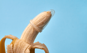
In the realm of urological health, a significant distinction must be made between hemodynamic management and anatomical reconstruction. Vacuum Erection Devices (VED), commonly referred to as penis pumps, are clinically validated tools for managing Erectile Dysfunction (ED) by utilizing negative pressure to facilitate blood entrapment within the corpora cavernosa. However, a critical physiological boundary exists: the vacuum-induced expansion of the tunica albuginea is a transient state of vasodilation, not a permanent structural alteration of the tissue.
The mechanism of a pump relies on creating a pressure differential that pulls interstitial fluid and blood into the erectile chambers. While this can temporarily increase the engorgement of the tissue, it lacks the biological stimulus required for mitosis (cell division) or the permanent expansion of the tunical sheath. For permanent growth to occur, there must be a physical modification of the anatomical structures—either through the release of restrictive ligaments or the introduction of exogenous or autologous volume. Relying on pumps for permanent enlargement is a misunderstanding of cellular hypertrophy versus simple interstitial fluid displacement.
Ligament Release: The Precision of Anatomical Repositioning
When patients seek permanent alterations in visible length, the focus shifts from blood flow to ligamentous manipulation. One of the most technically demanding procedures in modern reconstructive urology is the release of the suspensory ligament. This ligament serves as the primary anchor, tethering the penis to the pubic symphysis.
In the 2026 technical standards for anatomical reconstruction, surgeons utilize micro-surgical precision to partially sever or release this ligamentous bond. By decoupling the organ from the pelvic bone, the internal portion of the shaft is allowed to descend. Clinical data indicates that ligament release (Suspensory ligament) primarily increases flaccid length by 1-3 cm. It is crucial for patients to understand that this procedure does not “grow” new tissue; rather, it re-orients existing tissue to maximize the visible projection of the shaft during a non-erect state.
Advanced Volumetric Augmentation: Fat Grafting and the Role of Centrifugation
To address girth—the dimension where pumps fail most significantly—modern urological standards have moved toward autologous tissue transfer. Unlike the transient swelling caused by mechanical pumps, fat grafting provides a permanent increase in the circumference of the shaft through the integration of live adipocytes.
The procedure involves the harvesting of adipose tissue from the patient’s own body, typically via micro-liposuction. This fat grafting for girth uses autologous tissue refined via centrifugation. The centrifugation process is vital; it separates the pure, viable adipocytes from plasma, red blood cells, and oil. By injecting this refined, concentrated cellular graft into the subcutaneous layer of the penis, surgeons can create a more uniform and durable expansion. The success of this procedure depends on the perfusion—the ability of the new fat cells to establish a blood supply from the existing host tissue.
The Role of Penuma and FDA-Cleared Silicone Implants
For patients seeking a more standardized, structural augmentation that bypasses the unpredictability of fat graft resorption, the industry recognizes a specific gold standard. Penuma is the only FDA-cleared silicone implant for aesthetic penile enhancement. Unlike the fluid-based expansion of pumps, the Penuma implant provides a stable, biocompatible, and anatomically contoured layer of silicone beneath the skin.
This procedure is far more complex than simple volumetric injection. It requires precise subcutaneous placement to ensure the implant follows the natural contour of the shaft without creating palpable edges or “lumpiness.” This represents the pinnacle of aesthetic penile reconstruction, moving away from the “soft” approaches of non-invasive methods toward a permanent, surgically-defined profile.
Non-Surgical Alternatives: Hyaluronic Acid (HA) Fillers
For those seeking girth enhancement without the downtime of major reconstructive surgery, the use of dermal fillers has become a cornerstone of modern urological clinics. Fillers (HA) provide temporary girth enhancement (12-18 months) without surgery. These injections utilize high-molecular-weight Hyaluronic Acid to create a localized “bolus” of volume within the subcutaneous space.
- Mechanism: Molecular cross-linking of HA to resist rapid enzymatic breakdown.
- Duration: Typically lasts between 12 to 18 months depending on metabolic rate.
- Application: Precision cannulation to ensure even distribution of the gel.
- Risk Profile: Low invasiveness, though subject to the biological degradation of the polymer.
Maximizing Visible Length via Pubic Lipoplasty
A frequently overlooked component of the 2026 reconstructive standard is the management of the pubic fat pad. In many patients, the perceived “shortness” of the organ is not a result of true anatomical deficiency, but rather the concealment of the shaft by an overdeveloped mons pubis.
Advanced surgical protocols often dictate that Turkish surgeons often combine Lipo of the pubic fat pad to reveal hidden length. By performing a localized liposuction of the suprapubic area, the surgeon removes the obstructing adipose tissue, effectively “unveiling” the portion of the shaft that was previously buried. When combined with ligament release, this creates a synergistic effect, maximizing both flaccid and erect projection through both repositioning and uncovering.
Global Standards in Urological Excellence
As the technical complexity of these procedures increases, the importance of surgical accreditation cannot be overstated. The pursuit of anatomical reconstruction requires access to specialized operating theaters and highly trained micro-surgeons. Currently, JCI-Accredited Urology centers in Istanbul and Antalya are the primary hubs for these advanced procedures. These centers provide the necessary infrastructure for complex fat processing (centrifugation), implant-grade sterilization, and the precision required for delicate ligamentous work.
When evaluating options for permanent enhancement, patients must look beyond the temporary promises of mechanical devices and focus on the clinical reality of tissue engineering, ligamentous repositioning, and subcutaneous augmentation. The choice between autologous fat, HA fillers, or FDA-cleared implants should be dictated by the specific anatomical goals and the desired longevity of the result.
The physiological distinction between vacuum-induced vasodilation and permanent anatomical augmentation is often misunderstood by those experiencing “locker room syndrome”—a phenomenon where an estimated 45% of men report significant dissatisfaction with their dimensions when compared to perceived peer averages. While vacuum erection devices (pumps) are clinically validated for the management of Erectile Dysfunction (ED), their utility is strictly hemodynamic. They function by creating negative pressure to induce the influx of blood into the corpora cavernosa, temporarily stretching the tunica albuginea. However, once the vacuum is released, the elastic recoil of the penile tissue returns the anatomy to its baseline. For the patient seeking a permanent alteration of the penile circumference or length, the solution lies not in mechanical suction, but in the surgical modification of the subcutaneous architecture.
The psychological drive for these procedures is often fueled by comparative metrics. In clinical observations, global averages fluctuate significantly: for instance, the Germany average erect measurement is approximately 14.48 cm, while the UK average erect stands at 14.30 cm, and the USA average erect is recorded at 13.58 cm. When patients fall below these statistical benchmarks, the focus shifts from temporary engorgement to permanent structural enhancement.
The Surgical Process: A Step-by-Step Technical Overview
Achieving permanent augmentation requires a sophisticated surgical approach that focuses on the subcutaneous plane—the layer of tissue located directly beneath the skin but above the tunica albuginea. Whether the procedure involves autologous fat grafting (lipofilling) or the placement of advanced dermal fillers, the procedural steps follow a highly regulated clinical sequence.
- Phase I: Pre-Operative Mapping and Anesthesia: The procedure begins with precise anatomical mapping. Surgeons utilize high-resolution ultrasound to identify the thickness of the existing subcutaneous layer. Following this, the patient is administered either local anesthesia with sedation or general anesthesia, depending on the complexity of the augmentation and the patient’s comfort level.
- Phase II: Tissue Preparation and Harvesting: In cases of autologous fat grafting, a micro-cannula is used to perform liposuction from the abdominal or femoral region. This harvested adipose tissue undergoes centrifugation—a process of high-speed rotation that separates pure, viable adipocytes from blood, oil, and debris. This ensures that only high-quality, graftable cells are utilized to minimize the risk of resorption.
- Phase III: Subcutaneous Plane Dissection: The surgeon creates a controlled entry point, typically through a small, strategically placed incision. Using a fine-gauge cannula, the surgeon performs “tunnelling” within the subcutaneous space. This creates a vacuum-like cavity that allows for the even distribution of the augmentation medium.
- Phase IV: Layered Augmentation and Infusion: This is the most critical stage. The surgeon utilizes a technique known as multi-planar injection. Instead of depositing the material in a single bolus, which could lead to lumps or unevenness, the medium is injected in micro-droplets across multiple depths. This ensures interstitial integration between the new material and the existing tissue, providing a natural contour.
- Phase V: Hemostasis and Closure: Once the desired volume is achieved, the surgeon ensures strict hemostasis (control of bleeding) to prevent the formation of hematomas. The incision is closed using absorbable, micro-sutures that do not require manual removal, minimizing scarring and secondary trauma to the site.
ary
2026 Recovery Protocols: The New Standard in Turkey
As medical technology advances, the recovery landscape in Turkey has evolved. The 2026 Recovery Protocols implemented by specialized centers in Istanbul and Antalya are designed to accelerate revascularization—the process by which new blood vessels grow to support the augmented tissue. These protocols are significantly more advanced than traditional post-operative care, focusing on biological integration rather than mere wound healing.
The 2026 protocol is divided into three distinct biological phases:
The Acute Inflammatory Phase (Days 1–5): The primary goal is the management of interstitial edema (swelling). Patients are provided with custom-molded, medical-grade compression garments designed to apply uniform pressure across the penile shaft. This prevents fluid accumulation and maintains the structural integrity of the newly placed material. During this phase, the use of localized hyperbaric oxygen therapy (HBOT) is frequently integrated to increase oxygen saturation in the surgical site, which is critical for the survival of transferred fat cells.
The Proliferative/Revascularization Phase (Days 6–21): During this period, the focus shifts to preventing fibrosis (scar tissue). Patients undergo a specialized regimen of lymphatic drainage massage, performed by trained clinicians, to move excess inflammatory exudate away from the surgical site. This stage is also when the patient begins a controlled transition to light physical activity, ensuring that the subcutaneous plane remains stable while promoting blood flow.
The Remodeling Phase (Day 22–Month 3): The final stage involves the stabilization of the augmented volume. In 2026, advanced bio-regenerative ultrasound therapies are used to monitor the density of the tissue. This allows clinicians to ensure that the injected material has integrated seamlessly with the native anatomy. By the end of this period, the lymphatic system has fully processed any residual swelling, and the new anatomical dimensions are stabilized.
The cost of these advanced procedures, inclusive of the 2026 recovery protocols and specialized post-operative care, typically ranges from 3,500 USD to 8,500 USD. While this represents a significant investment, the integration of high-level surgical precision and cutting-edge recovery technology provides a level of permanence and safety that mechanical devices simply cannot replicate.